 Dr. Harsh Vardhan Puri
 Call Us :
Hospital Address :
Medanta - The Medicity
CH Baktawar Singh Rd, Medicity, Islampur Colony, Sector 38, Gurugram, Haryana 122001
CH Baktawar Singh Rd, Medicity, Islampur Colony, Sector 38, Gurugram, Haryana 122001
Dr. Harsh Vardhan Puri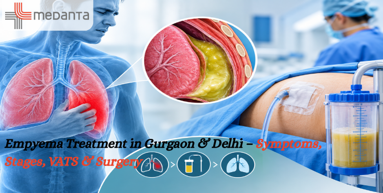
26 September, 2026
adminEmpyema is a serious condition in which infected fluid or pus collects in the pleural space, the area between the lung and the inner chest wall. It commonly develops as a complication of pneumonia or another infection and may interfere with normal lung expansion.
Timely diagnosis and appropriate treatment are important because untreated empyema can progress from a relatively simple fluid collection to a more organized infection with thick scar tissue around the lung.
For patients searching for Empyema Treatment in Gurgaon or Empyema Treatment in Delhi, evaluation by an experienced thoracic surgeon can help determine whether the condition can be managed with antibiotics and drainage or requires a surgical procedure such as VATS.
Dr. Harsh Vardhan Puri, Senior Consultant Thoracic & Lung Transplant Surgeon at Medanta – The Medicity, Gurugram, has specialized expertise in thoracic surgery and VATS key-hole surgery for empyema.
Empyema is the accumulation of infected fluid or pus in the pleural space surrounding the lung.
It most commonly develops following a chest infection such as pneumonia, but it can also occur with tuberculosis, chest trauma, infected pleural fluid, or after certain chest procedures and surgeries.
As infected fluid accumulates, the lung may not be able to expand normally. In more advanced cases, a thick fibrous layer can form around the lung, resulting in a condition commonly described as a trapped lung.
Empyema can develop due to several conditions, including:
The underlying cause is important because treatment may need to address both the infection and the infected pleural collection.
The symptoms can vary depending on the stage and severity of the infection.
Common symptoms include:
Patients who develop persistent breathing difficulty, high fever, worsening chest pain, or significant weakness after pneumonia should seek medical evaluation.
A detailed clinical evaluation is generally followed by appropriate imaging and laboratory investigations.
A chest X-ray can help identify fluid collection and changes in lung expansion.
A CT scan provides more detailed information about the pleural space, fluid collection, loculations, lung expansion, and surrounding structures.
Fluid from the pleural space may be collected for laboratory examination. Testing can help identify infection and guide antimicrobial treatment.
Depending on the individual case, additional blood tests, ultrasound, microbiological testing, and other investigations may be recommended.
Empyema generally progresses through three stages. Recognizing the stage helps the treating team determine the most appropriate treatment strategy.
In the early stage, infected or inflammatory fluid begins accumulating in the pleural space.
Depending on the patient’s condition, treatment may involve appropriate antibiotics and drainage.
Early treatment can help prevent progression to more complicated stages.
As the infection progresses, the fluid becomes thicker and fibrin can form multiple pockets or loculations.
These loculations can make simple drainage more difficult.
In selected patients, chest tube drainage, antimicrobial treatment, and additional interventions may be required. VATS can be considered when the infected collection cannot be adequately cleared with simpler treatment.
In advanced empyema, a thick fibrous peel can form around the lung.
This restrictive layer can prevent the lung from expanding properly and may result in a trapped lung.
In appropriately selected patients, surgical decortication may be required to remove the restrictive tissue and allow the lung to expand.
The treatment of empyema depends on its stage, severity, cause, patient’s general health, and response to initial treatment.
Patients searching for Empyema Treatment in Gurgaon may require one or more of the following approaches:
Antibiotics are an important part of treating the underlying infection.
The choice of antimicrobial treatment depends on the suspected or identified organism, severity of infection, laboratory findings, and individual patient factors.
Antibiotics alone may not be sufficient when a significant amount of infected fluid or pus has accumulated.
A chest tube may be inserted into the pleural space to drain infected fluid.
This can help remove the collection and allow the lung to expand.
The duration of drainage varies depending on the patient’s response and the amount and nature of the fluid.
When empyema becomes complicated or loculated, Video-Assisted Thoracoscopic Surgery (VATS) may be considered.
VATS is a minimally invasive thoracic surgical technique performed through small incisions using a camera and specialized instruments.
During the procedure, the surgeon can:
Medanta specifically lists Dr. Harsh Vardhan Puri’s expertise in Key-Hole (VATS) Surgery for Empyema.
In advanced or organized empyema, a thick fibrous peel may restrict lung expansion.
A procedure called decortication may be required in appropriately selected patients. The goal is to remove the restrictive fibrous tissue and allow the lung to expand more effectively.
Decortication can be performed using a minimally invasive approach in selected cases, while open surgery may be required in some advanced or technically difficult situations.
Patients searching for Empyema Treatment in Delhi may require specialist thoracic evaluation, particularly when the condition is recurrent, complicated, loculated, or not responding adequately to initial treatment.
Delhi NCR patients may also consider specialized thoracic surgical care in Gurugram.
Dr. Harsh Vardhan Puri provides thoracic and lung transplant surgical care at Medanta – The Medicity, Gurugram, including VATS treatment for empyema.
For patients travelling from Delhi, Noida, Faridabad, Ghaziabad, and other parts of NCR, the appropriate treatment location should be selected based on the patient’s medical requirements and the availability of specialized thoracic care.
Not every patient with empyema requires surgery.
Surgical treatment may be considered when:
The treatment decision should be made after reviewing the patient’s clinical condition and imaging findings.
VATS has become an important minimally invasive approach for selected patients with complicated empyema.
Instead of using a large chest incision, the surgeon uses small incisions to introduce a camera and specialized instruments.
Potential advantages of minimally invasive surgery may include:
However, VATS is not appropriate for every patient. Advanced disease, extensive scarring, previous surgery, anatomy, and the patient’s overall condition can influence the surgical approach.
Some patients with advanced or organized empyema may require open surgery.
When the pleural peel is very thick or the disease cannot be adequately treated using minimally invasive techniques, open decortication may be considered.
The objective remains the same: clear the infected material, remove restrictive tissue when necessary, and help the lung achieve better expansion.
Delaying treatment can allow the infection to become more organized and difficult to drain.
Early evaluation may help identify the condition before extensive pleural scarring develops.
Patients recovering from pneumonia who continue to have:
should discuss these symptoms with their treating doctor.
Dr. Harsh Vardhan Puri is a Senior Consultant Thoracic & Lung Transplant Surgeon at Medanta – The Medicity, Gurugram.
His listed areas of expertise include:
His Medanta profile specifically lists Key-Hole (VATS) Surgery for Empyema among his areas of specialization.
Medanta also describes VATS and robotic-assisted thoracic surgery as advanced approaches used for complex chest diseases, including advanced empyema.
Empyema is a serious infection that requires appropriate medical treatment. If left untreated, it can progress and may affect normal lung expansion.
Some patients can be managed with antibiotics and drainage, particularly when the condition is identified early. More complicated or organized empyema may require surgical treatment.
VATS may be considered when infected fluid is loculated, drainage is inadequate, or surgical clearance of the pleural infection is required.
Decortication is a surgical procedure used to remove a restrictive fibrous layer around the lung in selected patients with organized empyema. This can help the lung expand.
The duration varies according to the severity of infection, stage of empyema, treatment required, and the patient’s response. Some patients require prolonged antibiotics and drainage, while surgical cases require postoperative recovery and follow-up.
Recurrence is possible, particularly when the underlying infection or pleural collection is not adequately treated. Appropriate drainage, antimicrobial treatment, follow-up, and management of the underlying cause are important.
A thoracic surgeon may be particularly important when empyema is complicated, recurrent, loculated, associated with persistent air or fluid collection, or not improving with initial treatment.
Empyema is more than a simple accumulation of fluid around the lung. It is an infection that can progress through different stages and may eventually restrict lung expansion.
Treatment can include antibiotics, chest tube drainage, and, when necessary, minimally invasive VATS surgery or decortication.
For patients searching for Empyema Treatment in Gurgaon or Empyema Treatment in Delhi, an early assessment by an experienced thoracic specialist can help determine the appropriate treatment approach.
Dr. Harsh Vardhan Puri, Senior Consultant Thoracic & Lung Transplant Surgeon at Medanta – The Medicity, Gurugram, has specialized expertise in VATS surgery for empyema and other complex thoracic conditions.
Category : Empyema , Empyema Treatment